Category: Uncategorized

  • Why Polycystic Ovaries Are Now Called Poly Endocrine Metabolic Syndrome

    Why Polycystic Ovaries Are Now Called Poly Endocrine Metabolic Syndrome

    The old name made many people look in the wrong direction. “Polycystic ovaries” sounds like a problem limited to the ovaries, or like a condition caused by multiple cysts. In real life, the condition is often much wider. It can involve hormones, insulin, skin, periods, ovulation, weight changes, fertility, mood, and long-term metabolic health.

    That is why many doctors and researchers now prefer language that reflects the full picture, such as Poly Endocrine Metabolic Syndrome. The newer name is meant to reduce confusion and show that this is not just an ovarian condition. It is a body-wide endocrine and metabolic disorder.

    The exact terminology may vary between clinics, guidelines, and countries. Many medical records still use PCOS, short for polycystic ovary syndrome. Still, the thinking behind the name change matters because it changes how the condition is understood, discussed, and treated.

    This article is for general health education only. It cannot replace a consultation with a qualified gynaecologist, endocrinologist, or healthcare professional.

    Eye-level view of a menstrual calendar beside a bowl of fruit and a glass of water

    The old name created confusion

    The term “polycystic ovaries” has two main problems.

    First, it makes the condition sound like it is only about the ovaries. Many people hear the word “ovary” and assume the issue is limited to reproductive health. That can lead them to focus only on periods, pregnancy, or ultrasound findings.

    Second, the word “cystic” can create fear. People may imagine dangerous cysts that need urgent removal. In many cases, the so-called “cysts” seen on ultrasound are actually small follicles. These are tiny fluid-filled sacs that contain immature eggs. They are not the same as harmful ovarian cysts.

    This naming problem has affected how people react to a diagnosis. Some common misunderstandings include:

    • Thinking the condition is present only if cysts appear on ultrasound
    • Assuming normal-looking ovaries rule it out
    • Believing it affects only fertility
    • Ignoring insulin resistance, cholesterol, and blood sugar risks
    • Delaying care because symptoms feel “separate” from each other

    The phrase polycystic ovaries focuses on one possible sign. It does not explain the full syndrome.

    What Poly Endocrine Metabolic Syndrome means

    The newer name tries to describe the condition more accurately. Each part of the term carries meaning.

    Poly means many. The condition can affect several systems at once.

    Endocrine refers to hormones. In this condition, hormones such as androgens, insulin, luteinising hormone, and others may be involved. Higher androgen activity can contribute to acne, excess facial or body hair, scalp hair thinning, and irregular ovulation.

    Metabolic refers to how the body handles energy, glucose, insulin, and fats. Many people with this syndrome have insulin resistance, where the body needs more insulin than usual to keep blood sugar controlled. This may increase the risk of type 2 diabetes, fatty liver, high triglycerides, and other metabolic concerns over time.

    Syndrome means a pattern of features that often occur together. It does not mean every person has the same symptoms. One person may mainly have irregular periods and acne. Another may have fertility concerns. Someone else may have normal periods but signs of insulin resistance and high androgen levels.

    That variation is exactly why the broader name helps. It tells both patients and clinicians to look beyond the ovaries.

    Close-up view of a simple anatomical ovary model held beside a glucose meter

    Why the change matters in day-to-day care

    A name is not just a label. It shapes the first question people ask.

    If the name points only to ovaries, the first question becomes, “Do I have cysts?” If the name shows the endocrine and metabolic nature of the condition, the questions become more useful.

    Those questions may include:

    • Are my periods regular?
    • Am I ovulating?
    • Do I have symptoms of excess androgens?
    • Is insulin resistance present?
    • Are my blood sugar and lipids healthy?
    • Do I need lifestyle treatment, medication, or both?
    • What are my long-term health risks?

    This wider approach can lead to better care. A person with irregular periods may need cycle protection. A person with acne or excess hair growth may need treatment for androgen-related symptoms. A person with insulin resistance may benefit from nutrition changes, physical activity, weight management if relevant, and sometimes medicines prescribed by a doctor.

    For people trying to conceive, the broader view also helps. Fertility is not only about the ultrasound image. It also depends on ovulation, hormone balance, body weight changes where relevant, insulin levels, thyroid function, prolactin, semen parameters, tubal health, age, and other factors.

    The older name often made the condition sound simpler than it is. The newer name encourages a more complete assessment.

    Why ultrasound alone is not enough

    Ultrasound can be helpful, but it is not the whole diagnosis.

    Some people have polycystic-looking ovaries on ultrasound but do not have the syndrome. This can happen in healthy adolescents or adults who have no symptoms and regular cycles.

    On the other hand, some people may have the syndrome even if the ultrasound does not clearly show polycystic ovarian morphology. Symptoms, hormone tests, menstrual history, and metabolic markers may still point towards the diagnosis.

    This is one reason the old name caused trouble. It placed too much attention on a scan finding.

    A careful evaluation may include:

    • Menstrual cycle history
    • Acne, excess hair growth, or scalp hair thinning assessment
    • Blood tests for androgen levels where needed
    • Blood sugar testing
    • Insulin resistance assessment when clinically relevant
    • Lipid profile
    • Thyroid and prolactin tests to rule out similar symptoms
    • Ultrasound

    In India, many people first seek help because of irregular periods, acne, weight gain, or difficulty conceiving. A scan may be part of the work-up, but it should not be treated as the only deciding factor.

    Wide-angle view of a peaceful home exercise corner with a yoga mat and water bottle

    The condition is not the same for everyone

    One of the most frustrating parts of this condition is that it does not follow one pattern.

    Some people are lean. Some gain weight easily. Some have heavy bleeding. Some miss periods for months. Some have visible facial hair growth. Some have very few skin symptoms. Some discover the condition during fertility testing. Others learn about it after blood sugar changes.

    This variety can make people feel dismissed. A person may be told, “You don’t look like you have it,” or “Your scan is normal, so nothing is wrong.” Both statements can be misleading.

    A better approach is to identify the person’s pattern.

    For example, care may focus on:

    Cycle health

    Irregular or very infrequent periods may need medical attention. Long gaps between periods can sometimes affect the uterine lining, so doctors may recommend treatment to regulate bleeding or protect the lining.

    Androgen symptoms

    Acne, excess facial or body hair, and scalp hair thinning can affect confidence and quality of life. These symptoms deserve proper treatment, not dismissal as cosmetic issues.

    Metabolic risk

    Blood sugar, insulin resistance, cholesterol, blood pressure, sleep, and liver health may need attention, especially when there is a family history of diabetes or heart disease.

    Fertility planning

    Many people with this syndrome can conceive, but some may need help with ovulation. Early guidance can reduce months of uncertainty.

    The name Poly Endocrine Metabolic Syndrome makes space for all these patterns.

    Lifestyle advice should be practical, not shame-based

    Lifestyle changes can help many people, especially when insulin resistance plays a role. But the advice needs to be realistic and respectful.

    Telling someone to “just lose weight” is not good care. Weight can be influenced by hormones, sleep, stress, appetite signals, genetics, medicines, and daily routine. Some people with this condition do not have weight gain at all.

    A more useful plan looks at habits that support metabolic health:

    • Regular meals with enough protein
    • More fibre from vegetables, dals, beans, fruits, and whole grains
    • Reduced intake of sugary drinks and frequent refined snacks
    • Strength training and walking, when possible
    • Better sleep timing
    • Treatment for sleep apnoea if symptoms suggest it
    • Stress management that feels realistic
    • Follow-up blood tests when advised

    Indian food can fit well into a health plan. The goal is not to remove all rice, roti, dosa, or traditional meals. The goal is to improve balance, portion size, protein, fibre, and consistency.

    For example, a meal of dal, sabzi, curd, salad, and a measured portion of rice or roti may support steadier energy better than a meal built mainly around refined carbohydrates. Small changes, repeated daily, often work better than extreme diets.

    Medical treatment depends on the main concern

    No single treatment fits everyone. A doctor will usually base treatment on symptoms, age, pregnancy plans, test results, and health risks.

    Common treatment areas include:

    Concern

    Possible medical approach

    Irregular periods

    Hormonal medicines or cyclic treatment, based on doctor advice

    Acne or excess hair growth

    Hormonal therapy, anti-androgen medicines, dermatology care

    Insulin resistance

    Lifestyle care and sometimes insulin-sensitising medicines

    Fertility concerns

    Ovulation induction and fertility evaluation

    Metabolic risk

    Monitoring blood sugar, lipids, blood pressure, and weight-related factors

    Medicines should be taken only after medical evaluation. This matters because symptoms similar to this syndrome can also occur with thyroid disorders, high prolactin, adrenal conditions, medication effects, and other hormonal problems.

    The best care does not stop at prescribing tablets. It includes explaining what is being treated, what to monitor, and when to review the plan.

    Overhead view of a balanced Indian meal with dal, vegetables, curd, and roti

    The new name can reduce stigma

    The word “ovary” in the old name often made the condition feel private, embarrassing, or limited to fertility. Many people stayed silent because they did not want to discuss periods, hair growth, acne, or conception plans.

    A broader name can make the conversation more medical and less judgemental. It helps explain that the condition involves hormones and metabolism, not personal failure.

    This matters for teenagers too. Adolescents may have irregular periods for normal developmental reasons in the first few years after menarche, so diagnosis needs care. At the same time, severe acne, excess hair growth, rapid weight changes, or long gaps between periods should not be ignored. The newer framing encourages doctors and families to look at the whole person rather than rush to label based only on ultrasound.

    Better language can also help partners and families understand the condition. When people hear “metabolic syndrome,” they may take nutrition, activity, sleep, and follow-up tests more seriously. When they hear only “ovarian cysts,” they may expect a quick scan-based answer.

    What has not changed

    The name may evolve, but the core facts remain.

    This is still a common hormonal condition. It can affect periods, skin, hair, fertility, and metabolic health. It still needs individualised care. Ultrasound still has a role in some cases. Blood tests and clinical history still matter. Long-term follow-up is still useful.

    Most of all, the condition is manageable. Many people improve symptoms, protect long-term health, and achieve pregnancy when desired with the right support.

    The main change is the lens. Instead of seeing the condition as “cysts in the ovaries,” the newer name asks everyone to see the larger pattern.

    The takeaway

    The shift from “polycystic ovaries” to Poly Endocrine Metabolic Syndrome is not just a change in wording. It corrects a misunderstanding.

    The older name made the condition sound like an ultrasound finding. The newer name better reflects the truth: this is a hormone and metabolism-related syndrome that may affect many parts of health.

    Clearer language can lead to better questions, earlier testing, kinder conversations, and more complete treatment. If symptoms such as irregular periods, acne, excess hair growth, scalp hair thinning, weight changes, or fertility concerns are present, the next step is not panic. The next step is a proper medical evaluation that looks beyond the ovaries.

  • Understanding Follicular Studies on Ultrasound for Enhanced Fertility Insights

    Understanding Follicular Studies on Ultrasound for Enhanced Fertility Insights

    Follicular studies on ultrasound have become a cornerstone in fertility evaluation and treatment. These studies provide detailed information about ovarian follicles, which are crucial for understanding ovulation and reproductive health. For individuals or couples facing fertility challenges, follicular monitoring offers a window into the body's natural cycle, helping healthcare providers tailor treatments and improve chances of conception.

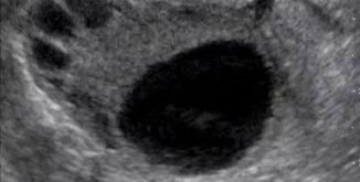
    Close-up ultrasound image of ovarian follicles during follicular study

    What Is a Follicular Study?

    A follicular study is a series of ultrasound scans performed during a woman’s menstrual cycle to track the growth and development of ovarian follicles. Follicles are fluid-filled sacs in the ovaries that contain immature eggs. Each cycle, several follicles begin to grow, but usually only one reaches full maturity and releases an egg during ovulation.

    The study typically starts early in the cycle, around day 8 to 10, and continues every two to three days until ovulation is confirmed. This monitoring helps identify the size, number, and quality of follicles, providing valuable information about ovulation timing and ovarian function.

    Why Follicular Studies Matter in Fertility

    Understanding follicular development is essential for diagnosing and managing fertility issues. Here’s why follicular studies are important:

    • Timing Ovulation: Knowing the exact day of ovulation helps couples time intercourse or insemination for the best chance of conception.
    • Assessing Ovarian Reserve: Follicular growth patterns can indicate how well the ovaries respond to hormonal signals, reflecting fertility potential.
    • Guiding Fertility Treatments: In assisted reproductive technologies like IVF or IUI, follicular monitoring helps determine the right time for egg retrieval or insemination.
    • Detecting Abnormalities: Irregular follicular development can signal hormonal imbalances, polycystic ovary syndrome (PCOS), or other reproductive disorders.

    How Follicular Studies Are Conducted

    The procedure is non-invasive and uses transvaginal ultrasound, which provides clear images of the ovaries and follicles. Here’s what to expect:

    1. Initial Scan: Performed early in the menstrual cycle to check baseline follicle size and uterine lining.
    2. Follow-up Scans: Conducted every 1-2 days to measure follicle growth and endometrial thickness.
    3. Ovulation Confirmation: When a follicle reaches about 18-24 mm, ovulation is expected soon. A final scan confirms follicle rupture or egg release.

    Each scan takes about 10-15 minutes and is generally painless. The ultrasound probe is inserted into the vagina, offering a close view of the ovaries.

    Interpreting Follicular Study Results

    The key measurements during a follicular study include:

    • Follicle Size: Mature follicles typically measure between 18 and 24 mm before ovulation.
    • Number of Follicles: Multiple mature follicles may indicate hyperstimulation or conditions like PCOS.
    • Endometrial Thickness: A lining thickness of 7-14 mm is considered optimal for implantation.
    • Follicle Growth Rate: Healthy follicles grow approximately 1.5-3 mm per day.

    Doctors use these parameters to decide the best timing for interventions or to adjust medications.

    Practical Examples of Follicular Study Use

    • Natural Cycle Monitoring: A woman with irregular periods undergoes follicular studies to pinpoint ovulation, improving natural conception chances.
    • Clomiphene Citrate Treatment: A patient taking ovulation-inducing drugs is monitored to ensure follicles respond appropriately and to avoid overstimulation.
    • Intrauterine Insemination (IUI): Follicular studies help schedule insemination when the follicle is mature.
    • In Vitro Fertilization (IVF): Multiple scans track follicle development to plan egg retrieval precisely.

    Factors Affecting Follicular Study Accuracy

    Several factors can influence the quality and interpretation of follicular studies:

    • Timing of Scans: Missing the optimal scan window can lead to inaccurate ovulation prediction.
    • Ultrasound Equipment: High-resolution machines provide clearer images.
    • Operator Skill: Experienced sonographers improve measurement reliability.

    Tips for Patients Undergoing Follicular Studies

    • Stay Consistent: Attend all scheduled scans for accurate monitoring.
    • Track Your Cycle: Note menstrual dates and symptoms to share with your doctor.
    • Ask Questions: Understand what each scan shows and how it affects your treatment.
    • Follow Instructions: Some clinics may advise on hydration or bladder fullness for better imaging.

    Considerations

    Tracking follicular growth optimizes timing but can not ensure successful conception or implantation.

    Additionally, follicular size alone does not guarantee egg quality or fertility success. Combining ultrasound findings with hormone tests and clinical evaluation offers a more complete picture.

    The Future of Follicular Monitoring

    Advances in ultrasound technology and artificial intelligence promise more precise and automated follicular assessments. Portable ultrasound devices and telemedicine may also increase accessibility for patients in remote areas.

    Tracking follicular development remains a vital tool in reproductive medicine, helping many individuals and couples understand their fertility and make informed decisions.

    Follicular studies on ultrasound offer a clear view of ovarian activity, guiding fertility treatments and natural conception efforts. By monitoring follicle growth and ovulation timing, healthcare providers can tailor approaches that improve outcomes. If you are exploring fertility options, discussing follicular monitoring with your doctor can provide valuable insights and support your journey toward parenthood.

  • Penile Doppler Ultrasound What It Is and Why It Matters

    Penile Doppler Ultrasound What It Is and Why It Matters

    Erectile problems are often treated as a private frustration. However, they can also indicate how well blood is moving through the body. This is why a test that examines penile blood flow can provide useful answers. This is especially true when tablets are ineffective, the cause is unclear, or surgery is being considered.

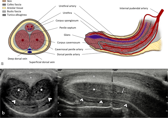
    A penile Doppler ultrasound is a medical imaging test that checks blood flow in the arteries and veins of the penis. It helps doctors understand whether erectile dysfunction is linked to poor arterial inflow, venous leakage, scarring, injury, or another physical cause.

    Eye-level view of an ultrasound machine beside a covered examination couch.

    What the Test Checks

    A standard ultrasound uses sound waves to create images of structures inside the body. Doppler ultrasound adds another layer. It shows the speed and direction of blood flow.

    For penile assessment, the test mainly looks at two things:

    • Arterial inflow

    This measures how well blood enters the penis through the cavernosal arteries.

    • Venous outflow

    This assesses whether blood stays trapped long enough to maintain an erection.

    An erection requires a precise sequence. Arteries widen, blood fills spongy tissue, and veins compress to keep blood inside. If the arteries do not supply enough blood, the erection may be weak. If veins allow blood to drain too quickly, the erection may not last.

    The scan can also reveal changes in tissue structure. This is particularly relevant for individuals with penile curvature, pain, trauma, plaques, or suspected fibrosis.

    Why Doctors Recommend It

    Not every individual with erectile dysfunction requires this scan. Many cases can be evaluated through medical history, physical examination, blood tests, and response to medications. A doctor may suggest Doppler imaging when the answer is less clear.

    Common reasons for recommending this test include:

    • Erectile dysfunction that does not respond well to oral medications
    • Erectile issues in younger patients where a vascular cause is suspected
    • History of pelvic injury, penile trauma, or surgery
    • A suspected venous leak
    • Curvature or pain suggestive of Peyronie’s disease
    • Planning before certain treatments or procedures
    • Assessment after priapism or prolonged erection
    • Checking blood flow in complex cases where psychological and physical factors overlap

    The results can help avoid guesswork. For example, two individuals may have similar symptoms but different causes. One may have reduced arterial blood flow due to diabetes, smoking, high blood pressure, or vascular disease. Another may have normal inflow but early drainage through veins. Their treatment plans may differ significantly.

    How the Procedure Usually Works

    The individual lies down in a private examination area. A clinician applies ultrasound gel to ensure the probe makes clear contact with the skin. Baseline images are taken first.

    In many cases, the doctor administers a small injection of a medication (papaverine) into the side of the penis. This medication helps relax blood vessels and produce an erection, allowing blood flow to be measured under stimulated conditions. The injection may sound concerning, but it uses a fine insulin syringe needle and is usually quick. Some individuals feel a brief sting or pressure. Oral sildenafil citrate with audiovisual stimulation techniques can also be used.

    The scan continues at timed intervals. The clinician measures blood flow in the penile arteries and checks whether veins are draining too much blood. The test typically takes around 20 to 60 minutes, depending on the protocol and how the body responds.

    Privacy should be maintained throughout the procedure.

    Close-up view of an ultrasound probe resting beside a tube of gel on a medical tray.

    What the Measurements Mean

    A report may include several technical terms. The treating doctor should explain them in plain language, but it is helpful to understand what they refer to.

    Peak Systolic Velocity

    This measures how strongly blood enters during the pumping phase of the heartbeat. Low values may suggest poor arterial inflow. This can occur with vascular disease, diabetes, high cholesterol, smoking-related blood vessel changes, or after certain injuries.

    End Diastolic Velocity

    This examines blood flow when the heart is resting between beats. If this remains high after an erection develops, it may indicate that blood is leaving too quickly through the veins.

    Resistive Index

    This is a calculated value that provides more context about resistance in the blood vessels. It is interpreted alongside other measurements, symptoms, and clinical findings.

    Structural Findings

    The scan may also describe plaques, scarring, calcification, or asymmetry. In Peyronie’s disease, ultrasound can help identify plaque location and whether it contains calcium. This information can influence treatment decisions.

    A single number rarely provides the full answer. Anxiety during the test, medication response, pain, hormone status, and other health conditions can all affect results. Good interpretation requires both the scan and the clinical picture.

    What to Expect Before and After the Scan

    Preparation is usually straightforward. The clinic may ask for a brief history of medications, allergies, heart conditions, blood pressure issues, and previous reactions to injection medications. It is important to mention blood thinners, bleeding disorders, or any history of prolonged erections.

    Before the scan, the doctor may inquire about:

    • When the erectile problem started
    • Whether erections occur during sleep or early morning
    • Response to tablets such as sildenafil or tadalafil
    • Diabetes, hypertension, cholesterol, thyroid disease, or heart disease
    • Smoking, alcohol use, and other lifestyle factors
    • Pelvic surgery, spinal injury, or trauma
    • Pain, curvature, or lumps

    After the test, most individuals can return to routine activities. If an injection was used, the clinic should provide instructions about what to do if the erection lasts longer than expected. A prolonged erection requires prompt medical attention, as untreated priapism can damage tissue.

    Mild bruising, tenderness, or swelling at the injection site can occur. Severe pain, heavy bleeding, fever, or an erection that does not resolve should be reported urgently.

    Wide-angle view of a private diagnostic room with a privacy screen and folded patient gown.

    Benefits of the Test

    The primary value of the scan is clarity. Erectile dysfunction can have multiple causes, and they often overlap. Stress, performance anxiety, relationship concerns, poor sleep, medications, hormone imbalance, nerve damage, and blood vessel disease may all contribute.

    Doppler imaging helps identify whether there is a measurable vascular issue.

    Its benefits include:

    • Better Diagnosis

    It can differentiate between inflow problems and possible venous leakage.

    • Treatment Planning

    Results may guide medications, injections, vacuum devices, counseling, lifestyle changes, or surgical options.

    • Support for Complex Cases

    It can assist when symptoms and routine tests do not align.

    • Assessment of Penile Tissue

    It may reveal plaques, scar tissue, or changes after injury.

    • Useful Follow-Up

    In selected cases, it can help track changes after treatment.

    For many patients, the test also reduces uncertainty. Having a physical explanation can make the next steps feel less vague.

    Limits and Possible Risks

    No test is perfect. Doppler results can be influenced by technique, medication dose, anxiety, pain, room temperature, and timing of measurements. An individual who feels very tense during the scan may not respond as they would in a relaxed setting.

    Possible risks are usually minor but should be discussed.

    These may include:

    • Temporary discomfort from the injection
    • Small bruise or swelling
    • Dizziness or flushing, depending on the medication used
    • Painful erection
    • Prolonged erection requiring treatment

    The rare but significant concern is priapism, an erection that lasts too long and does not resolve. Clinics that perform this test should have a plan to manage it.

    The scan also does not replace a full health check. Erectile dysfunction can be an early sign of cardiovascular risk. Blood pressure, blood sugar, cholesterol, weight, sleep, and mental health all deserve attention.

    How Results Can Guide Treatment

    The report does not treat the condition by itself. Its role is to assist the doctor in choosing a sensible path.

    If arterial inflow is reduced, the focus may include vascular risk reduction. This can involve better diabetes control, blood pressure management, cholesterol treatment, cessation of tobacco use, exercise, weight management, and reviewing medications that may affect erections. Oral ED medications may still help some individuals, while others may require different options.

    If venous leakage is suspected, tablets may be less reliable. A doctor may discuss injection therapy, vacuum erection devices, constriction rings, or specialist procedures in selected cases.

    If the scan reveals plaques or scarring, treatment may depend on pain, curvature, sexual function, and whether the condition is stable or changing. Peyronie’s disease has various approaches, from observation and medications to procedures in selected patients.

    If blood flow appears normal, that result is also valuable. It may shift attention towards hormone levels, nerve function, medication side effects, anxiety, depression, relationship factors, sleep issues, or pelvic floor concerns.

    Questions to Ask Before Booking

    A brief conversation before the appointment can make the test less stressful. Useful questions include:

    • Who will perform and interpret the scan?
    • Will an injection be used?
    • Which medication is used for the injection?
    • What should be done if the erection lasts too long?
    • How long will the appointment take?
    • When will the report be ready?
    • Should any medications be paused before the test?
    • What follow-up is needed after the report?

    A scan should ideally be conducted at a center experienced in penile vascular studies, as technique and interpretation are crucial.

    The Takeaway

    Penile Doppler ultrasound is significant because it transforms a difficult symptom into measurable information. It can indicate whether blood flow is entering effectively, leaving too quickly, or affected by tissue changes. This can guide treatment more accurately than trial and error alone.

    The best next step is not to self-diagnose based on symptoms or search results. If erectile dysfunction is persistent, painful, sudden, linked with curvature, or not improving with treatment, I encourage you to speak with a qualified urologist. A careful assessment can protect both sexual health and overall well-being.

  • Understanding Ultrasound Findings in Polycystic Ovarian Syndrome

    Understanding Ultrasound Findings in Polycystic Ovarian Syndrome

    Polycystic ovarian syndrome (PCOS) is a common hormonal disorder affecting many women of reproductive age. One of the key tools doctors use to diagnose and monitor PCOS is ultrasound imaging. Understanding what ultrasound findings mean in PCOS can help patients and healthcare providers make informed decisions about treatment and management.

    Close-up view of a transvaginal ultrasound showing multiple small ovarian follicles

    What Is Polycystic Ovarian Syndrome?

    PCOS is a condition characterized by hormonal imbalance, irregular menstrual cycles, and often, multiple small cysts on the ovaries. These cysts are actually immature follicles that have not developed properly. Women with PCOS may experience symptoms such as:

    • Irregular or absent periods
    • Excess hair growth
    • Acne
    • Weight gain
    • Difficulty conceiving

    The exact cause of PCOS is not fully understood, but it involves a combination of genetic and environmental factors.

    Role of Ultrasound in Diagnosing PCOS

    Ultrasound imaging is a non-invasive method that uses sound waves to create pictures of the ovaries and uterus. It helps doctors see the size, shape, and structure of the ovaries and identify the presence of cysts or follicles.

    There are two main types of ultrasound used in PCOS diagnosis:

    • Transvaginal ultrasound: A probe is inserted into the vagina to get a close and clear view of the ovaries. This is the preferred method for detailed ovarian imaging.
    • Transabdominal ultrasound: The probe is placed on the abdomen. This method is less detailed but may be used in some cases.

    Key Ultrasound Findings in PCOS

    When examining an ultrasound for PCOS, doctors look for specific features that suggest the condition. These include:

    1. Increased Number of Follicles

    One hallmark of PCOS is the presence of many small follicles arranged around the edge of the ovary. Typically, these follicles measure 2 to 9 millimeters in diameter. The diagnostic criteria often mention:

    • 12 or more follicles in each ovary
    • Follicles arranged in a "string of pearls" pattern

    This pattern reflects the arrested development of follicles that do not mature properly.

    2. Enlarged Ovarian Volume

    Women with PCOS often have larger ovaries compared to those without the condition. An ovarian volume greater than 10 cubic centimeters is considered a significant finding. Enlarged ovaries result from the accumulation of multiple follicles and increased stromal tissue.

    3. Increased Ovarian Stromal Echogenicity

    The ovarian stroma is the connective tissue inside the ovary. In PCOS, this tissue often appears denser or brighter on ultrasound images. This increased echogenicity is linked to hormonal changes and may contribute to the symptoms of PCOS.

    4. Absence of Dominant Follicle

    Normally, during a menstrual cycle, one follicle becomes dominant and grows larger to release an egg. In PCOS, this process is disrupted, and no dominant follicle is seen on ultrasound. This explains the irregular or absent ovulation common in PCOS.

    How Ultrasound Findings Support Diagnosis

    The Rotterdam criteria, widely used for diagnosing PCOS, include ultrasound findings as one of three key features. A diagnosis requires two of the following:

    • Irregular or absent ovulation
    • Clinical or biochemical signs of excess androgens (male hormones)
    • Polycystic ovaries on ultrasound

    Ultrasound findings alone do not confirm PCOS but provide strong evidence when combined with symptoms and blood tests.

    Practical Tips for Patients Undergoing Ultrasound

    If you are scheduled for an ultrasound to check for PCOS, here are some tips to prepare:

    • Schedule the ultrasound early in your menstrual cycle, ideally between days 2 and 5, for the most accurate assessment.
    • Empty your bladder if you are having a transvaginal ultrasound to improve image quality.
    • Discuss your symptoms and medical history with your healthcare provider before the scan.
    • Ask for a copy of the ultrasound report to keep for your records and future consultations.

    Limitations of Ultrasound in PCOS

    While ultrasound is valuable, it has some limitations:

    • Not all women with PCOS have polycystic ovaries on ultrasound. Some may have normal-looking ovaries but still have the syndrome.
    • Polycystic ovaries can appear in women without PCOS. This means ultrasound findings must be interpreted alongside clinical symptoms.
    • Operator skill and equipment quality affect results. A skilled technician and high-quality machine provide clearer images.

    Advances in Ultrasound Technology

    New ultrasound techniques are improving the detection and understanding of PCOS:

    • 3D ultrasound offers detailed images of ovarian structure and volume.
    • Doppler ultrasound measures blood flow in the ovarian tissue, which may differ in PCOS.
    • Automated follicle counting software helps provide more accurate and consistent follicle counts.

    These advances may lead to better diagnosis and personalized treatment plans.

    What Ultrasound Findings Mean for Treatment

    Ultrasound results help guide treatment decisions. For example:

    • If many small follicles are present but no dominant follicle, ovulation induction medications may be recommended.
    • Enlarged ovaries with increased stromal tissue may indicate a need for hormonal therapy to regulate cycles.
    • Monitoring ovarian response during fertility treatments relies heavily on ultrasound imaging.

    Regular ultrasound follow-up can track changes in ovarian appearance and response to therapy.

    Summary

    Ultrasound plays a crucial role in identifying the characteristic ovarian changes seen in polycystic ovarian syndrome. Key findings include multiple small follicles, enlarged ovarian volume, and increased stromal echogenicity. These features, combined with clinical symptoms and hormone tests, help confirm the diagnosis and guide treatment.

    If you suspect you have PCOS or have been diagnosed, understanding your ultrasound results can empower you to participate actively in your care. Always discuss your findings with your healthcare provider to develop a plan tailored to your needs.

    Taking the next step means scheduling regular check-ups and following recommended treatments to manage symptoms and improve quality of life.

  • Understanding Thyroid Health and Ultrasound Screening

    Understanding Thyroid Health and Ultrasound Screening

    Thyroid health is crucial for overall well-being. The thyroid gland plays a significant role in regulating metabolism, energy levels, and hormone balance. Understanding how to monitor thyroid health can lead to early detection of potential issues. This is where ultrasound screening comes into play.

    What Is Thyroid Screening by Ultrasound?

    Thyroid screening by ultrasound is a non-invasive imaging technique that uses sound waves to create detailed pictures of the thyroid gland. The thyroid is a butterfly-shaped gland located in the neck that regulates metabolism, energy, and hormone balance. Ultrasound helps doctors examine the gland’s size, shape, and texture without radiation or discomfort.

    Unlike blood tests that measure hormone levels, ultrasound provides a visual assessment. It can detect nodules, cysts, inflammation, or other abnormalities that may not cause symptoms initially. This makes it a valuable tool for early diagnosis and monitoring.

    Why Is Thyroid Screening Important?

    Thyroid disorders are common and can range from harmless to serious. Some of the most frequent conditions include:

    • Thyroid nodules: Small lumps that may be benign or cancerous.
    • Goiter: Enlargement of the thyroid gland.
    • Thyroiditis: Inflammation due to autoimmune diseases or infections.
    • Thyroid cancer: Though rare, early detection improves treatment outcomes.

    Many people with thyroid problems do not experience obvious symptoms until the condition progresses. Screening by ultrasound can identify issues early, allowing timely intervention and reducing the risk of complications.

    Who Should Consider Thyroid Ultrasound Screening?

    Not everyone needs routine thyroid ultrasound screening. However, certain groups benefit from it, such as:

    • Individuals with a family history of thyroid disease or thyroid cancer.
    • People with a visible or palpable lump in the neck.
    • Patients with abnormal thyroid blood test results.
    • Those exposed to radiation in the neck area during childhood or adulthood.
    • Individuals with symptoms like unexplained weight changes, fatigue, or voice changes.

    Doctors decide on screening based on risk factors and clinical findings. If you have concerns about your thyroid health, discussing ultrasound screening with your healthcare provider is a good step.

    How Does the Ultrasound Procedure Work?

    The ultrasound procedure is simple, painless, and usually takes 15 to 30 minutes. Here’s what to expect:

    1. Preparation: No special preparation is needed. You may be asked to remove jewelry or clothing around the neck.
    2. Positioning: You lie on your back with your neck slightly extended.
    3. Gel application: A water-based gel is applied to the neck to help the ultrasound probe make good contact.
    4. Scanning: The technician moves a small handheld device called a transducer over your neck. It sends sound waves that bounce off the thyroid and create images on a monitor.
    5. Image capture: The technician captures images of the thyroid from different angles.
    6. Completion: The gel is wiped off, and you can resume normal activities immediately.

    The procedure does not use radiation and is safe for all ages, including pregnant women.

    What Can Ultrasound Detect in the Thyroid?

    Ultrasound can reveal a variety of thyroid conditions by showing detailed images of the gland’s structure:

    • Nodules: Ultrasound can measure size, shape, and composition (solid or cystic). It helps distinguish benign from suspicious nodules.
    • Cysts: Fluid-filled sacs appear as dark areas on the scan.
    • Calcifications: Tiny calcium deposits may indicate malignancy risk.
    • Inflammation: Changes in texture or blood flow suggest thyroiditis.
    • Enlargement: The gland’s overall size can be assessed to detect goiter.
    • Lymph nodes: Nearby lymph nodes can be examined for enlargement or abnormalities.

    Doctors use ultrasound findings along with clinical data to decide if further tests, such as biopsy or blood work, are needed.

    Benefits of Ultrasound Screening for Thyroid Health

    Ultrasound screening offers several advantages:

    • Non-invasive and painless: No needles or radiation involved.
    • Real-time imaging: Immediate visualization of the gland.
    • High sensitivity: Detects small nodules and subtle changes.
    • Guidance for biopsy: Helps target suspicious areas accurately.
    • Monitoring: Tracks changes over time without risk.

    These benefits make ultrasound a preferred first step in evaluating thyroid concerns.

    Limitations of Thyroid Ultrasound

    While ultrasound is powerful, it has some limitations:

    • It cannot determine if a nodule is cancerous without biopsy.
    • It does not measure thyroid hormone function.

    Therefore, ultrasound is part of a broader diagnostic approach, combined with clinical evaluation and lab tests.

    What Happens After the Ultrasound?

    After the scan, a radiologist reviews the images and sends a report to your doctor. The report may include:

    • Description of thyroid size and texture.
    • Details about any nodules or abnormalities.
    • Recommendations for follow-up or biopsy if needed.

    Your doctor will discuss the results with you and suggest next steps. Many nodules are benign and require only periodic monitoring. Suspicious findings may lead to fine-needle aspiration biopsy to confirm diagnosis.

    Tips for Patients Considering Thyroid Ultrasound

    If you are scheduled for thyroid ultrasound screening, keep these tips in mind:

    • Wear comfortable clothing with easy neck access.
    • Inform the technician about any neck surgeries or radiation exposure.
    • Ask questions about the procedure and what to expect.
    • Follow up promptly on results and recommendations.
    • Maintain regular check-ups if you have known thyroid conditions.

    Being informed helps reduce anxiety and ensures you get the most from the screening.

    The Future of Thyroid Ultrasound Screening

    Advances in ultrasound technology continue to improve thyroid screening. New techniques like elastography measure tissue stiffness, helping differentiate benign from malignant nodules. Artificial intelligence is also being developed to assist in image interpretation, increasing accuracy and consistency.

    These innovations promise earlier detection and better outcomes for thyroid patients.

    Conclusion

    In conclusion, understanding thyroid health and the importance of ultrasound screening is essential for early detection and effective management of thyroid disorders. By being proactive and informed, individuals can take charge of their health and ensure they receive the best care possible. If you have concerns about your thyroid health, consider discussing ultrasound screening with your healthcare provider.

    For more information on ultrasound services, visit Diagnopro Ultrasound Clinic.

  • The Critical Role of Near Accurate Cervical Length Measurement in Pregnancy Outcomes

    The Critical Role of Near Accurate Cervical Length Measurement in Pregnancy Outcomes

    Pregnancy is a complex journey where every detail matters, especially when it comes to monitoring the health of both mother and baby. One crucial factor that often determines pregnancy outcomes is the length of the cervix. Measuring cervical length with near accuracy can provide vital information that helps healthcare providers predict and prevent complications such as preterm birth. This post explores why precise cervical length measurement is essential, how it impacts pregnancy management, and what methods ensure the best results.

    Close-up ultrasound image showing cervical length measurement in pregnancy

    Why Cervical Length Matters in Pregnancy

    The cervix acts as a gatekeeper during pregnancy, holding the fetus inside the uterus until it is time for delivery. Its length and firmness are indicators of how well it can maintain pregnancy. A shorter cervix can signal a higher risk of premature labor, which is a leading cause of neonatal complications and infant mortality worldwide.

    • Short cervix increases risk of preterm birth: Research shows that women with a cervical length less than 25 millimeters before 24 weeks of gestation have a significantly higher chance of delivering prematurely.
    • Early detection allows timely intervention: Identifying cervical shortening early enables doctors to recommend treatments such as progesterone therapy or cervical cerclage to reduce the risk of early labor.
    • Improves pregnancy monitoring: Regular and accurate cervical length measurements help track changes over time, guiding decisions on hospitalization or activity restrictions.

    How Near Accurate Measurement Influences Care

    Near accurate cervical length measurement means obtaining results that closely reflect the true length of the cervix with minimal error. This precision is critical because even small inaccuracies can lead to misclassification of risk and inappropriate management.

    • Avoids unnecessary interventions: Overestimating cervical shortening might lead to unnecessary treatments, causing stress and potential side effects.
    • Prevents missed diagnosis: Underestimating cervical shortening can delay crucial interventions, increasing the risk of preterm birth.
    • Supports personalized care: Accurate data allows healthcare providers to tailor monitoring and treatment plans based on individual risk profiles.

    Methods for Measuring Cervical Length

    Several techniques exist for measuring cervical length, each with varying degrees of accuracy and practicality.

    Transvaginal Ultrasound(Gold standard)

    This method is considered the gold standard for cervical length measurement due to its high accuracy and reproducibility.

    • Procedure: A small ultrasound probe is gently inserted into the vagina to get a clear view of the cervix.
    • Advantages: Provides precise images, less affected by maternal body habitus or bladder filling.

    Transabdominal Ultrasound

    This non-invasive method uses an ultrasound probe on the abdomen.

    • Procedure: The probe is placed on the lower abdomen to visualize the cervix.
    • Advantages: More comfortable for the patient.
    • Limitations: Less accurate due to interference from bladder filling, fetal position, and maternal obesity.

    Best Practices for Accurate Measurement

    To achieve near accurate cervical length measurement, healthcare providers should follow these guidelines:

    • Use transvaginal ultrasound when possible: It offers the most reliable results.
    • Ensure proper patient preparation: The bladder should be empty to avoid distortion of the cervix.
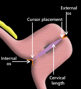
    • Standardize measurement technique: Measure the shortest functional length of the cervix from the internal to the external os.
    • Repeat measurements if necessary: Confirm findings with multiple scans to rule out errors.
    • Train sonographers thoroughly: Skilled operators reduce variability and improve accuracy.

    Impact of Accurate Cervical Length Measurement on Pregnancy Outcomes

    Accurate cervical length measurement directly influences clinical decisions that improve pregnancy outcomes.

    • Reduces preterm birth rates: Studies indicate that screening high-risk women and treating those with short cervices can lower preterm birth by up to 50%.
    • Improves neonatal health: Preventing early delivery reduces complications such as respiratory distress syndrome, infections, and long-term disabilities.
    • Optimizes resource use: Targeted interventions avoid unnecessary hospital stays and treatments, benefiting healthcare systems and families.
      Funneling of cervix-A structural change in the cervix during pregnancy where the internal opening (internal os) begins to dilate and open up prematurely, while the external opening (external os) remains tightly closed.. This ca
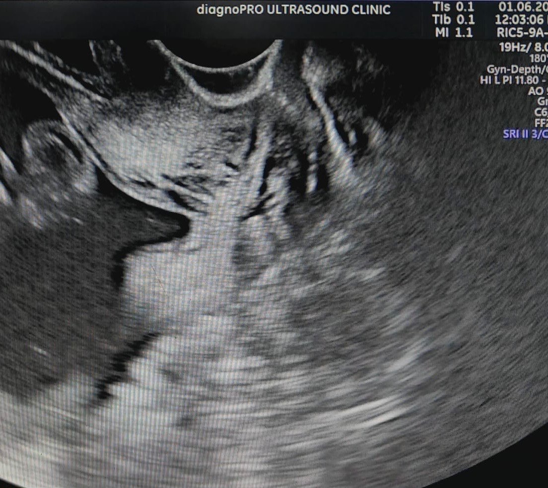
    • A woman with a history of preterm birth. At 20 weeks, a transvaginal ultrasound at our ultrasound clinic reveals her cervical length is 22 millimeters, below the 25-millimeter threshold with cervical funneling. Greater than 50% funneling before 25 weeks is associated with an 80% risk of preterm delivery.Timely management is of utmost importance.
    TVS ultrasound revealing short cervical length with cervical funneling
    • We followed up a previous case of cervical incompetence whose cervical length was 24mm and her gynacologist had done cervical encerclage ,had advised patient for rest , medical treatment and close ultrasound monitoring.The healthy baby was delivered at around 36weeks.

    RADIOLOGIST/SONOLOGIST ERRORS-

    Applying too much probe pressure,lack of experience, misidentifying the internal os, using the wrong ultrasound approach, and failing to record the shortest measured value.

    Radiologist or sonographer errors in measuring cervical length usually stem fro

    Future Directions in Cervical Length Measurement

    Advances in technology and research continue to improve the accuracy and accessibility of cervical length assessment.

    • Automated ultrasound analysis: Software algorithms can assist in measuring cervical length, reducing human error.
    • Portable ultrasound devices: These allow for wider screening in low-resource settings.

    Summary

    Near accurate measurement of cervical length plays a critical role in predicting and preventing preterm birth. It guides timely interventions, supports personalized care, and improves outcomes for mothers and babies. Healthcare providers should prioritize precise measurement techniques, especially transvaginal ultrasound, to ensure reliable data. Pregnant women, particularly those at high risk, benefit from regular cervical length monitoring as part of comprehensive prenatal care.

    Taking steps to improve cervical length measurement accuracy can make a meaningful difference in pregnancy outcomes. If you are pregnant or care for pregnant women, discuss cervical length screening with your healthcare provider to understand its role in your pregnancy journey.

  • The Importance of Doppler Ultrasound in Pregnancy

    The Importance of Doppler Ultrasound in Pregnancy

    What Is Doppler Ultrasound and How Does It Work?

    Doppler ultrasound is a specialized type of ultrasound that uses sound waves to measure the movement of blood through vessels. Unlike a standard ultrasound that creates images of the baby and uterus, Doppler ultrasound focuses on the flow of blood in the umbilical cord, placenta, and fetal heart.

    The device emits high-frequency sound waves that bounce off moving red blood cells. The changes in the sound waves, known as the Doppler effect, allow the machine to calculate the speed and direction of blood flow. This information helps doctors assess how well oxygen and nutrients are reaching the baby.

    Why Doppler Ultrasound Is Important During Pregnancy

    Monitoring blood flow is critical because it reflects the baby's health and development. Here are some key reasons why Doppler ultrasound is used:

    • Detecting Placental Problems

    The placenta supplies oxygen and nutrients to the baby. If blood flow through the placenta is reduced, it may indicate issues such as placental insufficiency, which can lead to growth problems.

    • Assessing Fetal Well-being

    Doppler ultrasound can check the blood flow in the umbilical artery and fetal brain. Abnormal flow patterns may signal fetal distress or conditions like intrauterine growth restriction (IUGR).

    • Monitoring High-Risk Pregnancies

    Women with conditions like high blood pressure, diabetes, or previous pregnancy complications often receive Doppler ultrasounds to closely track fetal health.

    • Guiding Delivery Decisions

    If Doppler results show poor blood flow, doctors may decide to induce labor or perform a cesarean section to protect the baby.

    When Is Doppler Ultrasound Typically Performed?

    Doppler ultrasound is not part of routine prenatal visits for all pregnancies. It is usually recommended in the second or third trimester when there is a medical reason to check blood flow. Common scenarios include:

    • Suspected fetal growth problems
    • High blood pressure or preeclampsia in the mother
    • Multiple pregnancies (twins or more)
    • Previous pregnancy complications
    • Reduced fetal movements reported by the mother

    The timing and frequency depend on the individual case and the healthcare provider’s judgment.

    Eye-level view of a pregnant woman undergoing Doppler ultrasound scan

    What to Expect During a Doppler Ultrasound Exam

    The procedure is safe, painless, and non-invasive. Here’s what typically happens:

    • The patient lies on an exam table.
    • A gel is applied to the abdomen to help transmit sound waves.
    • The doctor moves a handheld device called a transducer over the belly.
    • The Doppler machine displays blood flow patterns and heartbeats on the screen.
    • The exam usually takes 10 to 20 minutes.

    Results are often reviewed immediately by the doctor, who will explain what the findings mean for the pregnancy.

    Understanding Doppler Ultrasound Results

    Doctors look for specific patterns in blood flow to evaluate fetal health:

    • Normal Blood Flow

    Indicates the baby is receiving enough oxygen and nutrients.

    • Increased Resistance

    May suggest the placenta is not functioning well, requiring closer monitoring.

    • Absent or Reversed End-Diastolic Flow

    A serious sign that blood flow is severely compromised, often leading to urgent medical decisions.

    It is important to remember that Doppler ultrasound is one part of a comprehensive prenatal assessment. Results are interpreted alongside other tests and clinical information.

    High angle view of Doppler ultrasound printout showing fetal heart rate and blood flow waveforms

    Safety and Limitations of Doppler Ultrasound

    Doppler ultrasound is considered safe when used appropriately by trained professionals. It uses sound waves, not radiation, so it does not harm the mother or baby. However, it should only be performed when medically indicated to avoid unnecessary exposure.

    Limitations include:

    • It cannot diagnose all fetal conditions.
    • Results may vary depending on the doctor's skill and equipment quality.
    • It is less useful in early pregnancy.

    Final Thoughts on Doppler Ultrasound in Pregnancy

    Doppler ultrasound provides a window into the baby’s health by measuring blood flow and heart function. It plays a vital role in managing high-risk pregnancies and detecting potential problems early. For expectant parents, understanding this tool can reduce anxiety and support informed discussions with healthcare providers.

    If your doctor recommends a Doppler ultrasound, it means they want to ensure the best care for you and your baby. Ask questions, share your concerns, and use the information to stay actively involved in your pregnancy journey. Staying informed helps you prepare for a healthy delivery and a strong start for your child.

    The Future of Doppler Ultrasound Technology

    As technology advances, the capabilities of Doppler ultrasound are expected to improve. New techniques and equipment may provide even more detailed information about fetal health. This could lead to earlier detection of potential issues and better outcomes for mothers and babies.

    Furthermore, ongoing research is likely to enhance our understanding of how blood flow affects fetal development. This knowledge can inform best practices in prenatal care, ensuring that expectant parents receive the most effective and timely interventions.

    In conclusion, Doppler ultrasound is an invaluable tool in prenatal care. It not only helps monitor the health of the fetus but also empowers parents with knowledge and reassurance during their pregnancy journey. By embracing this technology, we can contribute to safer pregnancies and healthier outcomes for families.

  • The Importance of Fetal Echocardiography in Prenatal Care

    The Importance of Fetal Echocardiography in Prenatal Care

    Understanding Fetal Echocardiography

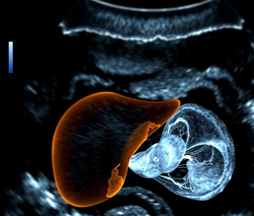
    Fetal echocardiography has emerged as a crucial diagnostic tool for the early identification of cardiac anomalies in developing fetuses. This sophisticated imaging modality enables healthcare professionals to evaluate the anatomical and functional integrity of the fetal heart. By doing so, it enhances the overall prognosis for both the mother and the infant.

    Key Benefits of Fetal Echocardiography

    Fetal echocardiography offers several key benefits:

    • Early Detection: This procedure identifies potential heart defects before birth, allowing for timely intervention.
    • Improved Outcomes: Early diagnosis can lead to better management strategies, improving the prognosis for affected infants.
    • Informed Decision-Making: It provides parents with crucial information about their baby's health, aiding in planning for delivery and postnatal care.
    • Specialist Referrals: The procedure facilitates early referral to pediatric cardiologists or specialized care teams when necessary.

    Indications for Fetal Echocardiography

    Fetal echocardiography is recommended in several situations:

    • Family history of congenital heart disease
    • Abnormal findings on routine ultrasounds
    • Maternal conditions such as diabetes or lupus
    • Increased nuchal translucency or other markers of potential abnormalities

    The Procedure: What to Expect

    During the fetal echocardiography:

    1. The mother is positioned on an examination table, and a gel is applied to her abdomen.
    2. An ultrasound transducer is positioned over the abdomen to capture images of the fetal heart.
    3. Multiple views of the heart are obtained to evaluate its structure, blood flow, and function.

    The procedure usually takes about 30 to 60 minutes. The mother may be asked to change positions to get better views of the heart. Images and measurements will be analyzed by a specialist.

    Interpreting Results

    The results of the fetal echocardiogram can indicate:

    • Normal heart development
    • Presence of congenital heart defects
    • Need for further evaluation or intervention

    Follow-Up Care

    If a congenital heart defect is detected:

    • The healthcare provider will discuss potential implications and management plans.
    • Additional tests or consultations with pediatric cardiologists may be recommended.

    Conclusion: The Role of Fetal Echocardiography

    Fetal echocardiography serves as a pivotal diagnostic tool in prenatal care. It offers invaluable insights into fetal cardiac development and facilitates the appropriate management of any identified abnormalities.

    Future Considerations in Fetal Echocardiography

    • Advancements in Technology: Ongoing improvements in imaging techniques and software may enhance the accuracy and detail of fetal echocardiography.
    • Training and Education: Continued education for practitioners in fetal echocardiography is vital to ensure high-quality care and interpretation of results.
    • Research: Further studies are needed to better understand the implications of various findings and to refine screening protocols.

    The Importance of Reliable Diagnostic Imaging

    In the realm of prenatal care, the significance of reliable diagnostic imaging cannot be overstated. It is essential for ensuring the health and well-being of both the mother and the developing fetus. As a patient, you deserve access to the best possible care, and that includes advanced imaging techniques like fetal echocardiography.

    By choosing a facility that prioritizes precision and reliability, you can feel confident in the care you receive. At Diagnopro Ultrasound clinic, we strive to be the go-to place for precise and reliable diagnostic imaging in Jammu & Kashmir. Our aim is to provide top-notch ultrasound services for all ages while maintaining high standards of care.

  • Is Fasting required for every ultrasound

    Is Fasting required for every ultrasound

    # Understanding Ultrasound Procedures: Fasting Requirements and Guidelines

    ## Fasting for Abdominal Ultrasounds

    Ultrasounds that typically necessitate fasting for 6 to 8 hours are abdominal scans (hepatobiliary). This preparation is crucial for optimal visualization of the gallbladder. The presence of food in the stomach can interfere with the clarity of the images obtained during the ultrasound procedure. When a patient fasts, the gallbladder, which stores bile produced by the liver, becomes more prominent and easier to visualize. This enhanced visibility allows the sonographer or radiologist to assess the gallbladder for any abnormalities such as gallstones, inflammation, or other potential issues that could affect its function.

    During the fasting period, it is advisable for patients to avoid not only solid foods but also liquids that may stimulate the digestive system, such as coffee or juices. Instead, clear water is typically permitted, as it helps to keep the body hydrated without affecting the gallbladder's status. Patients should also be informed about the importance of adhering to the fasting guidelines to ensure the highest quality of imaging results.

    In addition to gallbladder assessment, fasting may also aid in the examination of adjacent structures in the hepatobiliary system, including the liver, bile ducts, and pancreas. By ensuring that the stomach is empty, the ultrasound can provide a clearer view of these organs, allowing for a comprehensive evaluation of any potential pathologies.

    ## Ultrasounds That Do Not Require Fasting

    No fasting is usually required for pelvic, obstetric, thyroid, breast, scrotal, KUB, vascular doppler, or musculoskeletal ultrasounds. This is an important consideration for patients preparing for these types of imaging studies, as fasting can often be a source of discomfort and inconvenience. In contrast to abdominal ultrasounds, where fasting is typically mandated to ensure that the stomach is empty and to enhance the clarity of the images obtained, these specific ultrasound examinations can be performed without the need for dietary restrictions.

    For instance, during a pelvic ultrasound, the focus is on the organs within the pelvic region, including the bladder, uterus, and ovaries. Patients are usually encouraged to drink water prior to the exam to ensure that the bladder is full, which can help in obtaining clearer images. Similarly, in obstetric ultrasounds, which are performed to monitor the development of a fetus during pregnancy, there is no requirement for fasting. Instead, the primary goal is to visualize the fetus and assess its health and growth, which can be effectively achieved without any dietary limitations.

    Thyroid ultrasounds, which are conducted to examine the thyroid gland for abnormalities such as nodules or enlargement, also do not necessitate fasting. This is advantageous for patients who may be on medication for thyroid conditions, allowing them to maintain their regular medication schedule without interruption. In the case of breast ultrasounds, which are often used as a complementary tool alongside mammography, there are no fasting requirements, making it easier for patients to schedule their appointments at their convenience.

    Furthermore, scrotal ultrasounds are performed to evaluate conditions affecting the male reproductive system, such as testicular torsion or varicoceles, and similarly do not require fasting. This ensures that patients can undergo the examination without the added stress of having to abstain from food or drink beforehand. For the KUB ultrasound, which focuses on the kidneys, ureters, and bladder, fasting is not necessary, allowing for a more comfortable experience for the patient.

    When it comes to vascular doppler ultrasounds, which assess blood flow in the veins and arteries, there is also no fasting required. This is particularly beneficial for patients who may have conditions that affect their vascular health, as it allows for timely evaluation and intervention without the delays associated with fasting protocols. Lastly, musculoskeletal ultrasounds, used to visualize muscles, tendons, and joints, also do not require fasting, making it easier for patients to receive prompt care for their injuries or conditions.

    ## The Importance of Understanding Fasting Guidelines

    In summary, the absence of fasting requirements for these various types of ultrasounds not only enhances patient comfort but also facilitates easier scheduling and adherence to medical appointments. This approach reflects a growing understanding of patient needs and the importance of making diagnostic procedures as accessible and convenient as possible.

    No fasting is required for any emergency ultrasound scans.

  • Fetal Ultrasound

    Fetal Ultrasound

    Understanding Fetal Ultrasound: A Comprehensive Guide

    Fetal ultrasound, also known as sonography, is a crucial medical imaging technique used during pregnancy. It provides expectant parents and healthcare providers with valuable insights into the development and health of the fetus. This blog will explore the types, benefits, and what to expect during a fetal ultrasound.

    What is Fetal Ultrasound?

    Fetal ultrasound uses high-frequency sound waves to create images of the developing fetus in the womb. This non-invasive procedure helps monitor the growth, anatomy, and overall health of the baby. It is a safe method that does not involve radiation, making it suitable for use throughout pregnancy.

    Types of Fetal Ultrasound

    There are several types of fetal ultrasound, each serving different purposes:

    1. Transabdominal Ultrasound

    This is the most common type of ultrasound performed during pregnancy. A gel is applied to the abdomen, and a transducer is moved over the skin to capture images of the fetus.

    2. Transvaginal Ultrasound

    Used primarily in early pregnancy, this method involves inserting a transducer into the vagina to obtain clearer images of the fetus and surrounding structures.

    3. 3D and 4D Ultrasound

    These advanced imaging techniques provide three-dimensional images and real-time video of the fetus, allowing parents to see detailed features and movements.

    Benefits of Fetal Ultrasound

    Fetal ultrasound offers numerous benefits, including:

    • Monitoring Development: Ultrasounds help track the growth and development of the fetus, ensuring it is progressing as expected.
    • Identifying Multiple Pregnancies: It can confirm whether a woman is carrying twins or more.
    • Detecting Abnormalities: Early detection of potential congenital anomalies or health issues can lead to better management and care.
    • Determining Gender: Many parents choose to find out the sex of their baby during the ultrasound.
    • Assessing Placental Health: Ultrasounds can evaluate the position and health of the placenta, which is crucial for a safe pregnancy.

    What to Expect During a Fetal Ultrasound

    Knowing what occurs during a fetal ultrasound can reduce any worries. Here's what you can anticipate:

    1. Preparation

    For a transabdominal ultrasound, you may be asked to drink water beforehand to fill your bladder, which can help provide clearer images.

    2. The Procedure

    You will lie on an examination table, and a gel will be applied to your abdomen. The technician will then move the transducer over your belly to capture images.

    3. Duration

    The procedure typically lasts between 20 to 60 minutes, depending on the type of ultrasound and the information needed.

    4. Results

    The technician may not provide immediate results, as a radiologist will analyze the images and share findings with your healthcare provider, who will discuss them with you.

    Conclusion

    Fetal ultrasound is an invaluable tool in prenatal care, offering insights that can help ensure the health and well-being of both mother and baby. By understanding the types, benefits, and procedures involved, expectant parents can feel more prepared and informed as they navigate their pregnancy journey. Always consult with your healthcare provider for personalized advice and recommendations regarding fetal ultrasound.